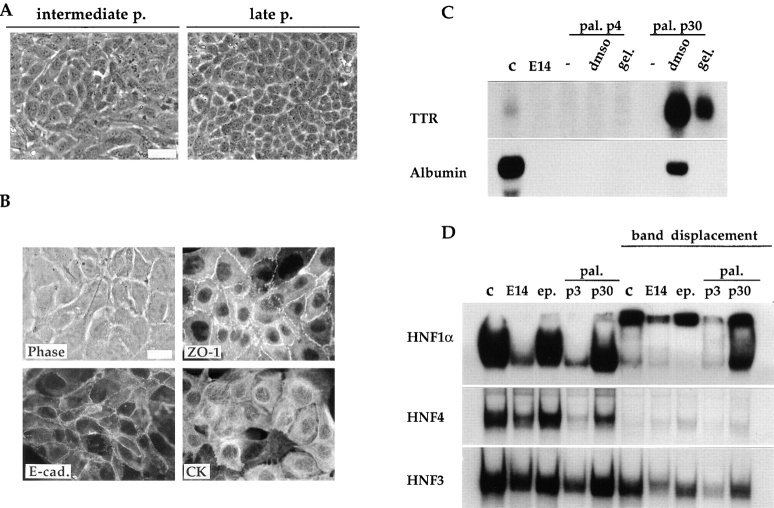
Figure 7.
Spontaneous phenotypic transition of the palmate cells toward an epithelial-hepatocytic phenotype. (A) Phase-contrast micrographs showing the morphology of the palmate cells at intermediate (p11) and late passages (p30). (B) Appropriate re-organization of the epithelial polarity markers (E-cad., ZO-1, and CK) in the palmate cells at passage 30. The phase-contrast and immunofluorescence micrographs of the ZO-1 visualization is of the same field. (C) Reexpression of hepatic functions after environmental treatments in the palmate cells late passage. Northern blot analysis of RNA extracted from the E14 parental line (E14), the palmate clone (pal.) at early (p4) and late (p30) passages as well as control Fao rat hepatoma cell line (c). Cultured cells were examined in the usual growth conditions (−), and 1 wk after addition of DMSO or gelatin. Each lane was loaded with 20 μg of total RNA. Probes used are indicated on the left. (D) Activation of LETF (HNF1α and HNF4) expression in palmate cells continuously kept in culture. The gel shift assays were performed with labeled oligonucleotides corresponding to the HNF1 site PE56, the HNF4 site C3P and the HNF3 site of the mouse TTR promoter, as detailed in Materials and Methods. The displacement of the protein-DNA complexes was obtained with anti-HNF1α or with an excess of unlabeled oligonucleotides for HNF4 and HNF3. Nuclear extracts of FGC4, as positive control (c), of E14 parental line (E14), of the epithelial (ep.) and palmate (pal.) clones at early (p3) and late (p30) passage were examined. Bar, 20 μm.

