
Figure 2.
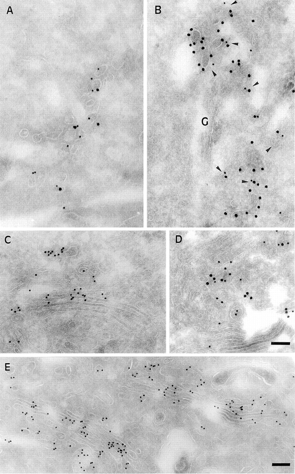
Kinetics of B-fragment transport from EE/RE to the Golgi apparatus revealed by immunoelectron microscopy in HeLa cells. (A) B-fragment was internalized for 1 h at 19.5°C, cells were fixed and prepared for cryosectionning as described under Materials and Methods. B-fragment (10-nm gold particles) was detected in tubular and vesicular elements that were also labeled for the TfR (15-nm gold particles). Cells that had internalized B-fragment at 19.5°C were then shifted for 2 min (B), 10 min (C–D), and 30 min (E) to 37°C. Cryosections were stained for B-fragment (10-nm gold particles in B, C, and E; 15-nm gold particles in D) and MPR46 (15-nm gold particles in B; 10-nm gold particles in D). Cryosections that were doubled stained for MPR46 (in B and D) showed that the B-fragment entered the Golgi apparatus via the TGN. Bars, 100 nm.