
Figure 1.
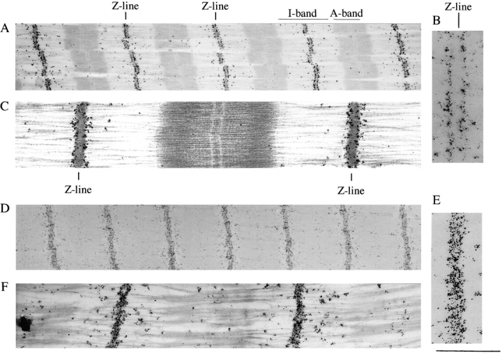
Immunoelectron microscopy of Z-disc titin. Fibers labeled with anti–titin Z1-Z2 antibodies (A–C). (A) Low-magnification image of a stained section. Darkly stained silver grains reveal epitopes at the edge of the Z-line. (B) High-magnification image of epitopes at edge of Z-line. Notice the absence of clear labeling inside the Z-disc. (C) High-magnification image of stained section. Fibers labeled with anti–titin Zr5-6 antibodies (D–F). (D) Low-magnification image of an unstained section. Darkly stained silver grains reveal epitopes throughout the width of the Z line. (E) High-magnification image. (F) High-magnification image of a stained section. Bar, 1 μm.