
Figure 10.
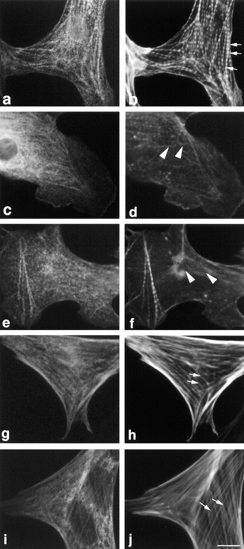
Microinjection of titin Z1-Z2 into the cytoplasm of cardiac myocytes results in disruption of myofibrils: stress fibers in fibroblasts are unaffected. Cells were microinjected with (a, b, g, and h) a nonspecific monoclonal antibody (MOPC-21) to identify injected cells and (c–f, i, and j) with the nonspecific antibody plus purified titin Z1-Z2. 2 h after injection, the cells were fixed and stained with (a) Cy2-conjugated anti–mouse antibodies and (b) Texas red–conjugated phalloidin. Note that the control microinjected antibody used to mark the injected cells appears to localize to some myofibrils in the cardiomyocytes and to some stress fibers in fibroblasts. Arrows point to the typical striated and stress fiber staining seen with Texas red–phalloidin (b) in cardiac myocytes and (h and j) in fibroblasts, respectively; arrowheads point to disrupted myofibrils in cardiac myocytes (d and f). Bar, 10 μm.