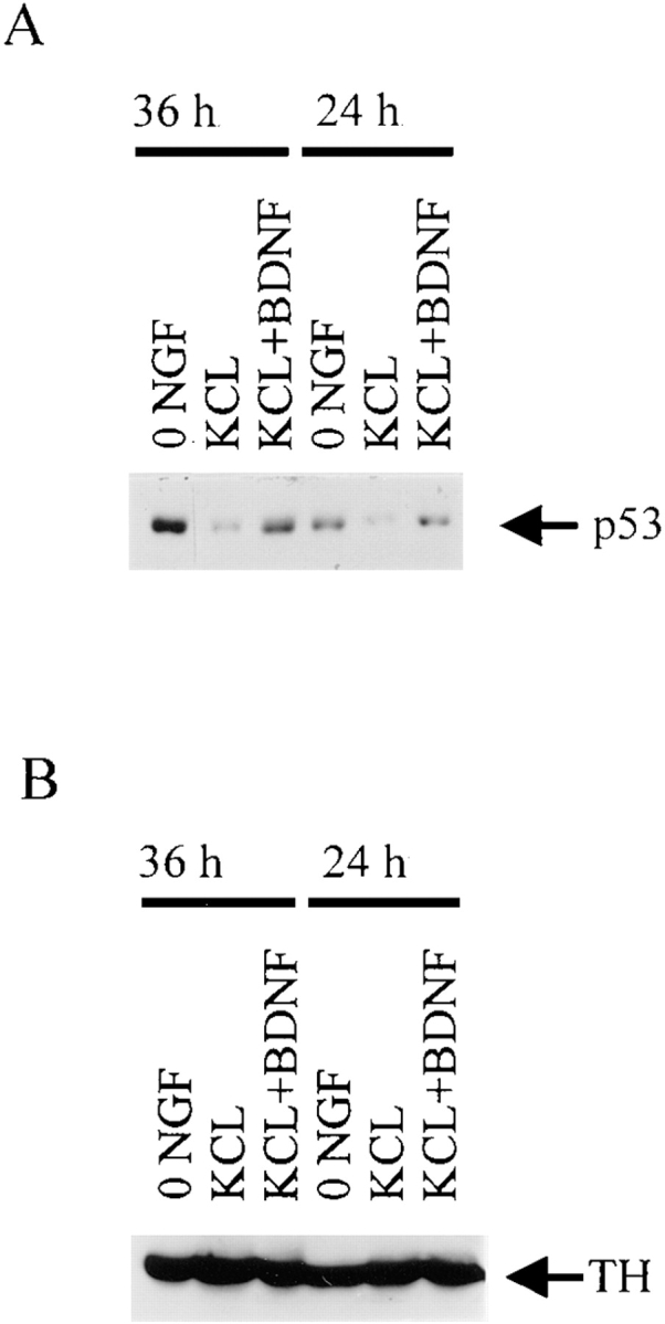
Figure 4.

p53 levels increase during apoptosis of sympathetic neurons as induced by BDNF-mediated activation of p75NTR. (A) Western blot analysis for p53 in equal amounts of protein derived from sympathetic neurons that were cultured in 50 ng/ ml NGF for 4 d, and then were washed free of NGF and switched into 50 mM KCl (KCL), 50 mM KCl plus 100 ng/ml BDNF (KCL + BDNF), or media containing no NGF or KCl (0 NGF) for 24 or 36 h. Note that p53 levels in the neurons treated with KCl and BDNF are similar to those in 0 NGF, and are greater than those maintained in KCl alone. (B) The same blot as in A reprobed for the neurotransmitter enzyme, tyrosine hydroxylase (TH) to demonstrate that equal amounts of protein were present in each of the lanes.
