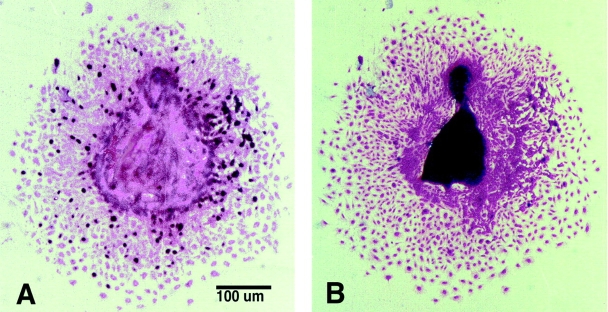
Figure 2.
Analysis of cell proliferation in neural crest outgrowth cultures. To quantitate cell proliferation in the neural crest outgrowth, BrdU incorporation was examined. Proliferating cells can be readily distinguished as those that are darkly stained after immunodetection with an anti-BrdU antibody (A). Such cultures were subsequently stained with hematoxylin for counting total cell number in the outgrowth (B). Note that A and B are pictures of the same outgrowth culture.

