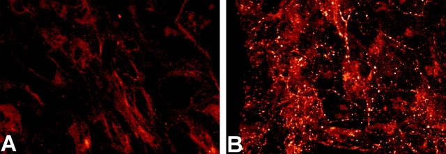
Figure 3.
Increased α1 connexin gap junction plaques in the CMV43 crest cells. Immunostaining with a α1 connexin antibody revealed increased α1 connexin expression in the homozygous CMV43 neural crest outgrowth (B) as compared with neural crest cells from nontransgenic embryos (A). The punctate pattern of immunostaining is consistent with the localization α1 connexins in gap junction plaques. In the control nontransgenic neural crest outgrowths, very small punctate immunostaining can be observed, although this is hard to record photographically.

