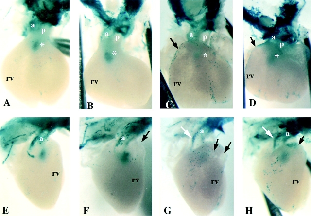
Figure 4.
Neural crest cells in fetal hearts of CMV43 and α1 connexin KO mice. The distribution of cardiac neural crest cells in E14.5 fetal hearts were examined using a lacZ reporter transgene driven by the α1 connexin promoter (Lo et al., 1997). Shown are the front and side views of X-gal–stained hearts from wild-type, CMV43, and α1 connexin KO mice. (A and E) Wild-type mouse heart. The white asterisk in A denotes presumptive neural crest cells in the conus, perhaps comprising cells in the closing seam of the aorticopulmonary septum. (B and F) Homozygous CMV43 heart. The black arrow in F denotes mild bulging of the conotruncus. Note a greater abundance of lacZ staining in the conus (B, white asterisk), suggesting an increased abundance of neural crest cells in the outflow septum. (C and G) Homozygous α1 connexin KO heart with severe conotruncal malformation. Note the very low level of lacZ staining in the conus (white asterisk), suggesting that fewer neural crest cells are present in the outflow septum. This heart exhibits a more severe phenotype consisting of very prominent thinning of the conotruncal myocardium (region denoted by black arrows). The white arrow in G denotes acute bend in aorta. (D and H) Homozygous α1 connexin KO heart with mild conotruncal malformation. This heart shows less prominent thinning of the conotruncal myocardium (region denoted by black arrows) as compared with the heart in (C and H). Interestingly, the level of lacZ staining in the conus is close to normal (D, white asterisk). The white arrow in H denotes an acute bend in the aorta. p, pulmonary trunk; a, aorta; rv, right ventricle.

