
Figure 10.
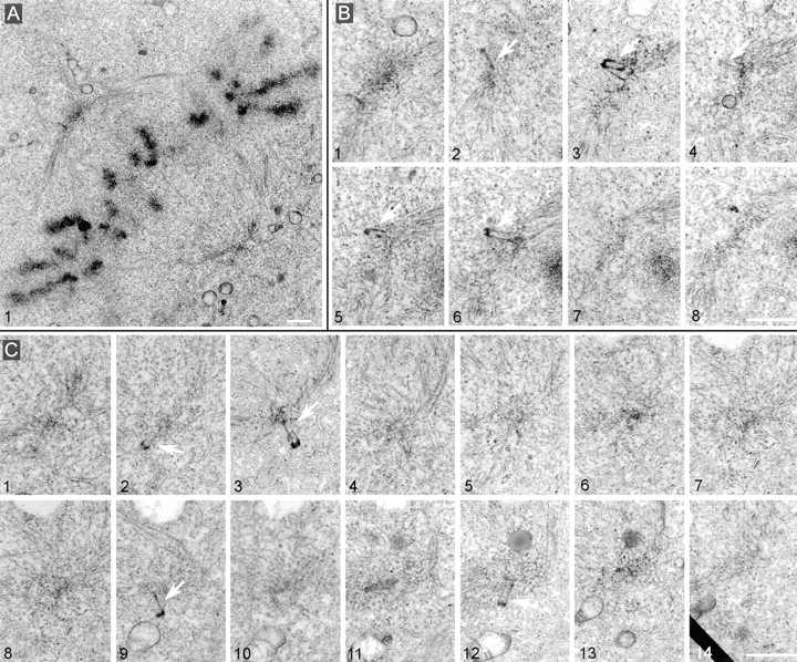
Metaphase spindles assemble in cells collected 60 h after loading with GT335 mAb but present partial centrioles at their poles. (A) Overview of the spindle. (B) Eight adjacent sections of the upper spindle pole. (C) 14 adjacent sections of the lower pole. Note in both cases the presence of several abnormal centrioles (arrows). They seemed excluded from the pericentriolar cloud and presented a weak staining of the walls. Pericentriolar material, however, was visible at both poles. Bars, 1 μm.