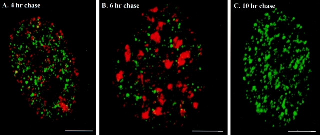
Figure 5.
Double labeling experiments in early S-phase versus later S-phase and the G2-phase. Replication sites in synchronized 3T3 fibroblasts were first labeled for 2 min with CldU (FITC secondary antibody, green sites), chased for 4–10 h and pulsed again for 5 min with IdU (Texas red secondary antibody, red sites). (A) 4-h chase (mid S-phase, type II for red RS); (B) 6-h chase (late S-phase, type III for red RS); (C) 10-h chase (G2-phase, there are no red RS). Bars, 5 μm.

