
Figure 1.
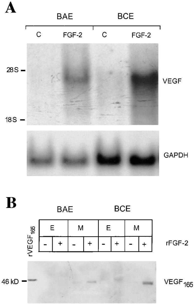
FGF-2 induces VEGF expression in endothelial cells. (A) Northern blotting analysis of total RNA (20 μg) from BAE or BCE cells treated with rFGF-2 (10 ng/ml) or with control medium (C) for 4 h. The RNA blot was hybridized with a DIG-labeled cDNA probe for human VEGF as described in Materials and Methods. GAPDH mRNA is shown as a control. The position of 28 S and 18 S ribosomal RNA is shown on the left. This experiment was repeated four times with comparable results. (B) Western blotting analysis of Triton X-100 extracts (E; 200 μg) or conditioned medium (M) from BAE or BCE cells treated with 10 ng/ml of rFGF-2 (+) or with control medium (−) for 17 h. The samples were electrophoresed under nonreducing conditions. The protein blot was hybridized with human VEGF IgG; antigen–antibody complexes were detected as described in Materials and Methods. Recombinant VEGF165 (10 ng) was run as a control in the leftmost lane. Molecular masses are shown in kD on the left. This experiment was repeated three times with comparable results.