
Figure 12.
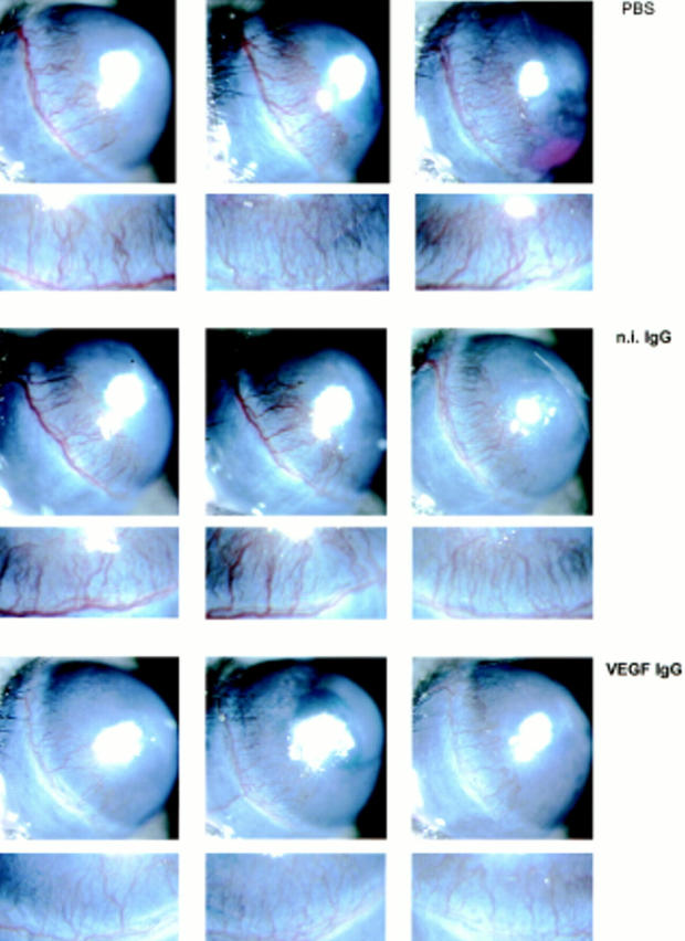
Effect of anti-VEGF antibody on FGF-2–induced angiogenesis. Hydron pellets containing 50 ng of rFGF-2 were implanted in the cornea of both eyes of 18 Swiss Webster mice as described in Materials and Methods. The animals were randomized into three groups of six mice and given i.v. injections of either PBS or PBS containing n.i. IgG (100 μg) or neutralizing anti-human VEGF monoclonal antibody (VEGF IgG; 100 μg) 1 d before pellet implantation and on postoperative days 1 and 3. The corneas were photographed by slit-lamp biomicroscopy on day 5 after implantation of the pellet. The eyes of the animals injected with VEGF antibody have fewer and thinner corneal limbic capillaries than those of animals injected with PBS or n.i IgG. An enlargement of the limbic area containing the newly formed vessels is shown below each photograph of the corresponding eye. This experiment was repeated twice with comparable results. In control mice that received corneal implants of pellets containing 200 ng of human recombinant VEGF, the same treatment with the VEGF antibody abolished the angiogenic response almost completely (data not shown).