
Figure 7.
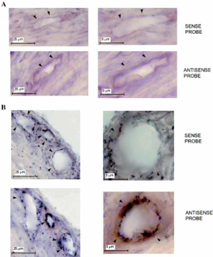
Expression of VEGF mRNA by vascular endothelium in vivo.In situ hybridization with DIG-labeled sense or antisense VEGF riboprobes of adjacent 30-μm sections of mouse corneas that received either (A) sham pellets or (B) pellets containing 50 ng of rFGF-2. Implantation of pellets in the cornea, preparation of the probes, and in situ hybridization were carried out as described in Materials and Methods. Contrast was enhanced by computer to increase the appearance of the reaction product. Arrowheads, vessel's wall. (A) Sections of limbic vessels show no hybridization with the probes. (B) Sections of newly forming capillaries in the stroma of the cornea show hybridization of the endothelium with the antisense but not with the sense probe. Hybridization signals (brown-black staining) are present only in the endothelium of newly formed vessels in FGF-2–treated eyes.