
Figure 9.
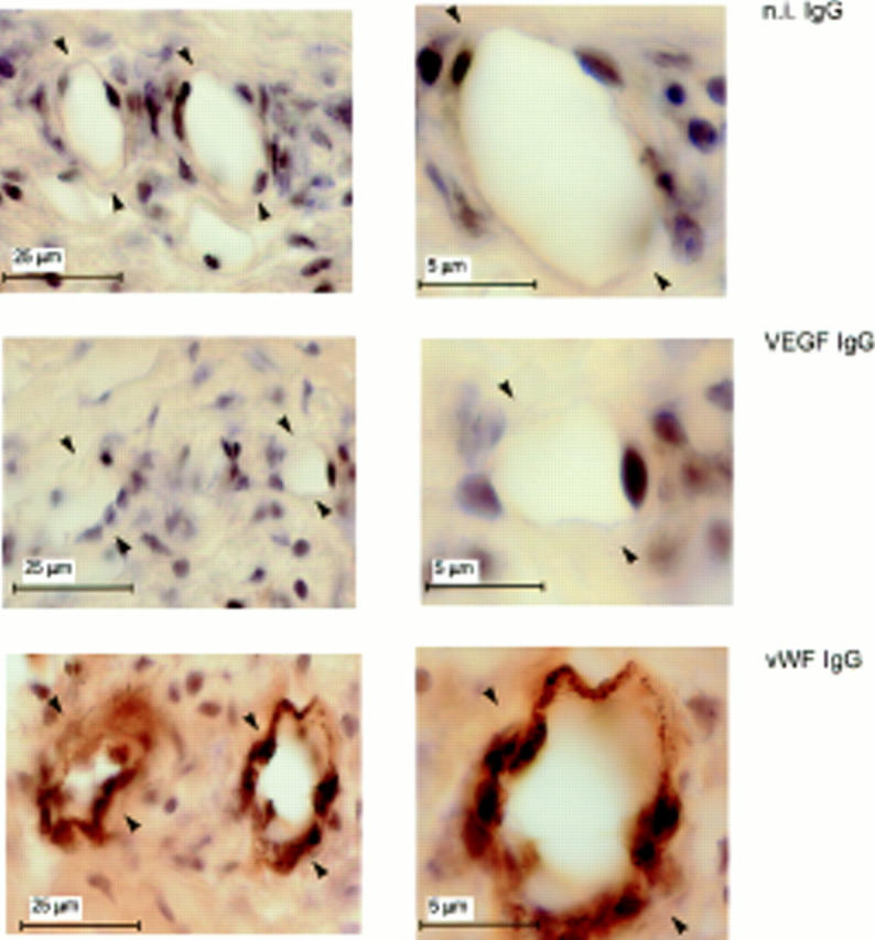
Lack of VEGF expression in quiescent endothelium. Adjacent 30-μm sections of mouse corneas that received pellets with no FGF-2 were immunostained with antibody to mouse VEGF (VEGF IgG) or to vWF (vWF IgG) or with n.i. IgG (n.i. IgG) as described in Materials and Methods. The endothelium of the limbic vessels (arrowheads) stains positively for vWF but not for VEGF.