
Figure 2.
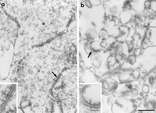

Behavior of the occludin and nine guanidine- insoluble bands on sonication followed by sucrose density gradient centrifugation. (a and b) Ultrathin sectional electron microscopic images of non-treated (a) and sonicated (b) junction fraction fixed in the presence of 0.1% tannic acid. The non-treated fraction was characterized by numerous actin filaments (*) and isolated junctions (arrow), which contained typical tight junctions (inset). After sonication followed by centrifugation, no actin filaments were detected, and isolated junctions were fragmented into small vesicular structures (arrow), some of which still contained typical tight junctions (inset). (c) Fractionation of sonicated junction fraction. After sonication, the isolated junctions were fractionated by stepwise sucrose density gradient centrifugation. 0:25%, 25:30%, 30:34%, 34:38%, 38:42%, and 42:50% interfaces were collected, and subjected to SDS-PAGE followed by silver staining. The distribution of the occludin band (occludin) was compared with those of nine guanidine-insoluble bands (band 1–9), and only band 9 was copartitioned with occludin. Silver staining and accompanying immunoblots with anti-occludin mAb (Oc-1) revealed that occludin was mainly recovered at 25:30%, 30:34%, and 34:38% interfaces, where band 9 was also characteristically accumulated. Bars in c indicate molecular masses of 200, 116, 97, 66, 45, 31, and 21 kD, respectively, from the top. Bar, 0.2 μm.