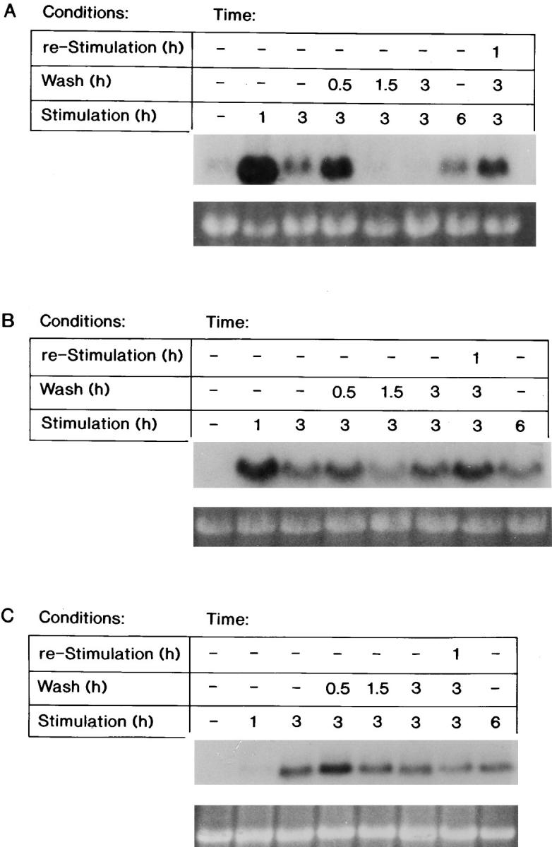
Figure 2.

The steady-state mRNA levels of FGF-1–induced genes after FGF-1 removal. RNA was extracted from Swiss 3T3 cells either at quiescence, after a 1-, 3-, or 6-h FGF-1 stimulation, or from populations that were transiently stimulated with FGF-1 (exposed to FGF-1 for 3 h, followed by a heparin wash and supplemented with DMI for 0.5, 1.5, or 3 h, or supplemented with DMI for 3 h and then reexposed to FGF-1 for 1 h). Northern blot analysis was performed as described in experimental procedures. Expression of the (A) Fos transcript (above), of the (B) Myc transcript (above), or of the (C) ODC transcript (above) with corresponding ethidium bromide–stained 18S rRNA (below) is shown.
