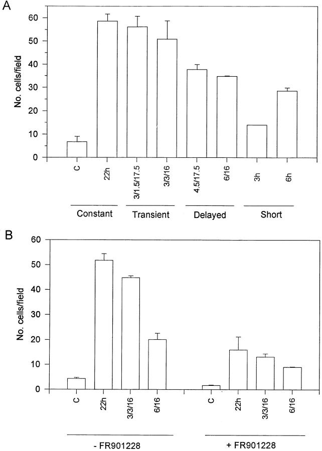
Figure 7.
(A) Migration of Balb/c 3T3 cells upon differential FGF-1 stimulation. Confluent monolayers of Balb/c 3T3 cells were wounded with a razor blade, and then stimulated with FGF-1 as described in Materials and Methods. Unstimulated cells were used as a comparative control (C). The number of cells migrating into the denuded area after a 22-h incubation at 37°C was determined by counting using a microscope with a grid at 100×. Because the total number of migratory cells varied between experiments, these data are the average of two independent experiments, two plates each, and are normalized to the level of total migration in the constantly-stimulated populations. Although the total number of migratory cells varied among experiments, the trends observed for the different populations were indeed consistent. (B) Migration of Balb/c 3T3 cells in the presence or absence of the Myc inhibitor FR901228. Confluent monolayers of quiescent Balb/c 3T3 cells were wounded and stimulated with FGF-1 in the presence of 2.5 ng/ml FR901228 as described in Materials and Methods. The various cell populations were stimulated in the absence of the inhibitor as a control.

