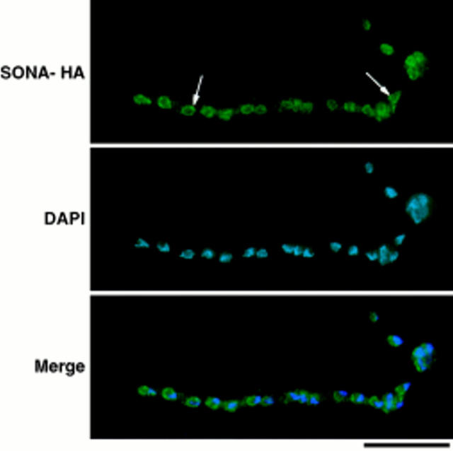
Figure 8.
SONA-HA localizes to the nuclear periphery. Cells of the SONA-HA–expressing strain, LPW42, were cultured, fixed, and then prepared for immunocytology as described in Materials and Methods. The image is identified to the left (SONA-HA shows 12CA5 staining). Arrows, example nuclei within the multinucleate cell shown. Digital images were captured using a Sensys Photometrics CCD camera and were merged using Phase 3 Imaging Systems software. Bar, 10 μm.

