
Figure 1.
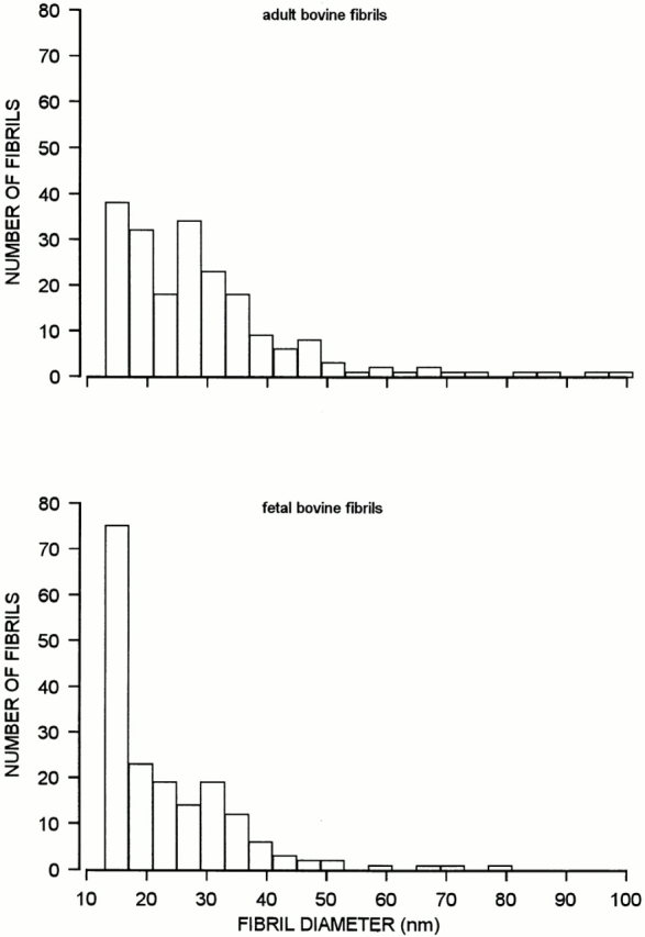
Histograms showing the diameter distribution of fibril fragments from bovine adult articular cartilage (top) and fetal cartilage (bottom), as determined by electron microscopy after negative staining. The fibril extracts were those described in the footnotes to Tables I and II. The overall number of measured fibrils were 202 and 181, respectively.