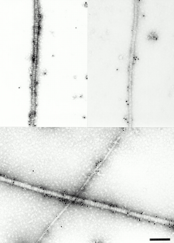
Figure 8.

Combined immunolocalization of decorin and collagen IX on fibril fragments from adult bovine articular cartilage. Rabbit anti-decorin antibodies were detected by 18-nm gold probes and mouse anti-collagen IX antibodies by 12-nm probes. Two representative examples of fibrils with diameters between 30 and 40 nm are shown (top). These fibrils, although weakly labeled (<5 gold particles per 10 D-periods), demonstrate colocalization of the two fibril constituents. A typical thin fibril carries dense label with collagen IX-probes only, whereas the thick 60-nm fibril is labeled exclusively for decorin (bottom). Bar, 200 nm.
