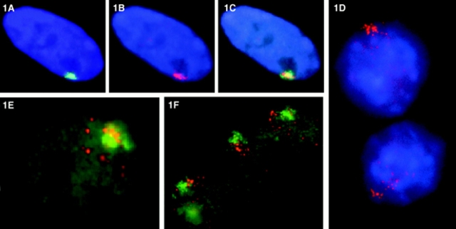
Figure 1.
Fluorescence in situ hybridization detection of XIST RNA in normal human and mouse/human hybrid cells. Digoxigenin and biotinylated probes were hybridized in situ and detected with fluorochrome-conjugated avidin or antidigoxigenin antibody. Fluorochromes used were FITC (green), rhodamine (red), and DAPI (blue). (A) In normal human diploid fibroblasts (46, XX WI-38 cells), the XIST RNA (green) occupies a discrete location in the nucleus. (B) The XIST RNA has a similar shape to the X chromosome territory defined by the whole X chromosome library signal (red; the weak red signal in the middle of this cell is the active X chromosome which is out of the plane of focus). (C) The XIST RNA essentially paints the X chromosome in these normal nuclei (overlap is yellow). (D) In mouse/human hybrids containing a single active human X chromosome (AHA-A5) that expresses XIST through treatment with the demethylating agent 5azadC, the XIST RNA (red) is disperse, and spreads throughout the nucleus. (E) The XIST RNA (red) does not associate or strictly localize to the human whole X chromosome library signal (green) in the hybrid cells. (F) In mouse/human hybrid cells (t86-B1maz1b-3a) that contain a single human inactive X that expresses XIST endogenously (no 5azadC treatment), the human XIST RNA (red) also shows aberrant localization to the whole X chromosome library signal (green).

