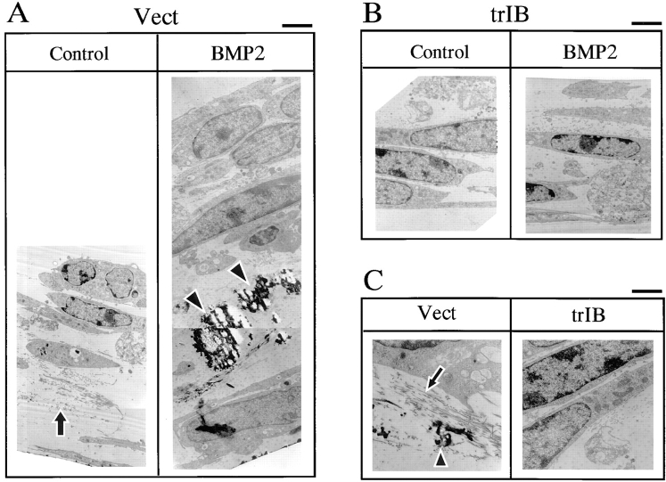
Figure 4.
Transmission electron microscopic analysis of the mineralized bone matrix structure of 2T3 cells. (A and B) Empty vector control (Vect) and trBMPR-IB (trIB)–expressing 2T3 clones were cultured for 12 d in the presence or absence of 100 ng/ml of BMP-2 and fixed with Na cacodylate and glutaraldehyde buffer for electron microscopy analysis. (C) High magnification of collagen fibers and mineralized bone matrix of 2T3 cells containing the empty vector (Vect) and trBMPR-IB (trIB). Arrows, unmineralized collagen fibers; arrowheads, mature mineralized matrix. Bars: (A and B) 2 μm; (C) 1 μm.

