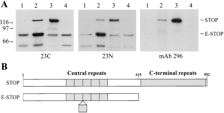
Figure 1.
STOP expression in DRG cells. (A) Immunoblot analysis of STOP expression. Proteins from 3-d differentiated DRG cells (lane 1), 10-d differentiated DRG cells (lane 2), adult rat brains (lane 3), and embryonic rat brains (lane 4) were run on 7.5% SDS gels. 20 μg of DRG cell extract proteins were loaded onto the gel. Amounts of loaded brain proteins were adjusted to equilibrate brain and DRG cell STOP signals. Proteins were immunoblotted with polyclonal STOP antibodies 23C and 23N (central repeat antibodies), and with monoclonal STOP antibody 296 (COOH-terminal repeat antibody) as indicated. The bands corresponding to STOP and E-STOP are indicated. Size markers are in kD. (B) Schematic representation of STOP and E-STOP showing domain structure of the two proteins. Both proteins contain a central domain composed of 5 (STOP) or 6 (E-STOP) repeats. STOP also contains a COOH-terminal repeat domain that is lacking in E-STOP. The E-STOP sequence ends at a position corresponding to aa 614 in the STOP sequence (Bosc et al., 1996). The E-STOP sequence data are available from GenBank/EMBL/DDBJ under accession no. AJ002556.

