Figure 3.
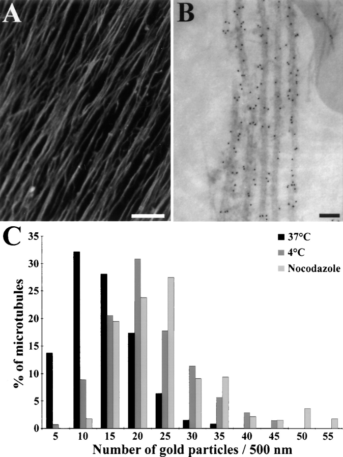
Immunolocalization of STOP proteins in DRG cell axons. After 10 d of culture, DRG cells were stained using the affinity-purified 23C STOP antibody and a Cy3- (A) or gold-labeled (B) secondary antibody. (A) Immunofluorescence analysis of STOP localization in DRG cell axons. Bar, 25 μm. (B) Immunoelectron microscopy of a DRG cell axon: microtubules were specifically decorated with gold particles. Bar, 50 nm. (C) Gold particle distribution along axonal microtubules in the distal part of DRG cell axons. (Black bars) total microtubules; (dark gray bars) cold stable microtubules; (light gray bars) nocodazole-resistant microtubules. In control experiments with secondary antibody alone, microtubules were never decorated with more than four gold particles.

