
Figure 2.
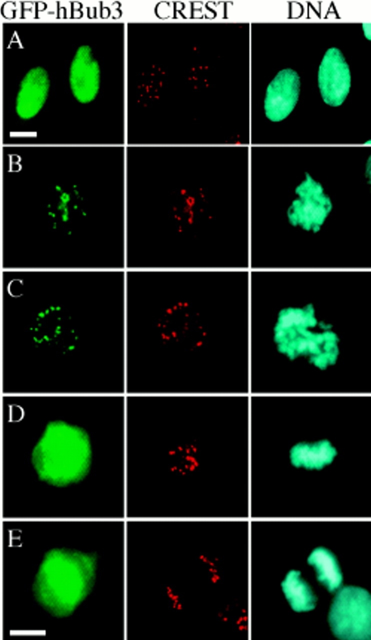
hBub3 localizes to kinetochores during prometaphase. BHK cells were transfected with a GFP–hBub3 expression construct, fixed, and then stained with a CREST antiserum to identify the kinetochores (red) and Hoechst dye to visualize the chromatin (blue). GFP fluorescence is shown in green. (A) Transfected cells showing that hBub3 is diffusely nuclear during interphase. (B) Transfected prophase and (C) prometaphase cells showing colocalization of GFP–hBub3 with kinetochores. (D) Transfected metaphase and (E) anaphase cells showing GFP–hBub3 diffusely distributed throughout the cell. Bars, 10 μm. B–D are to the same scale as E.