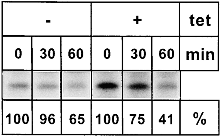
Figure 10.
Overexpression of the KKAA construct delays ER exit of procathepsin C. KKAA cells were cultured in presence or absence of tet for 48 h and pulse-labeled with [35S]methionine and chased for the indicated times. The cells were homogenized, and membranes were separated by Nycodenz gradient centrifugation (see Materials and Methods). Fractions 1–5 containing the ER marker p63 were pooled and Triton X-100 solubilized, and procathepsin C was immunoprecipitated. The immunoprecipitates were separated by SDS-PAGE, and procathepsin C was quantified from the fluorogram. The start signal after the pulse was set to 100%. All the lanes of this figure originate from the same fluorogram and were identically exposed. They had to be rearranged for reproduction purposes.

