
Figure 4.


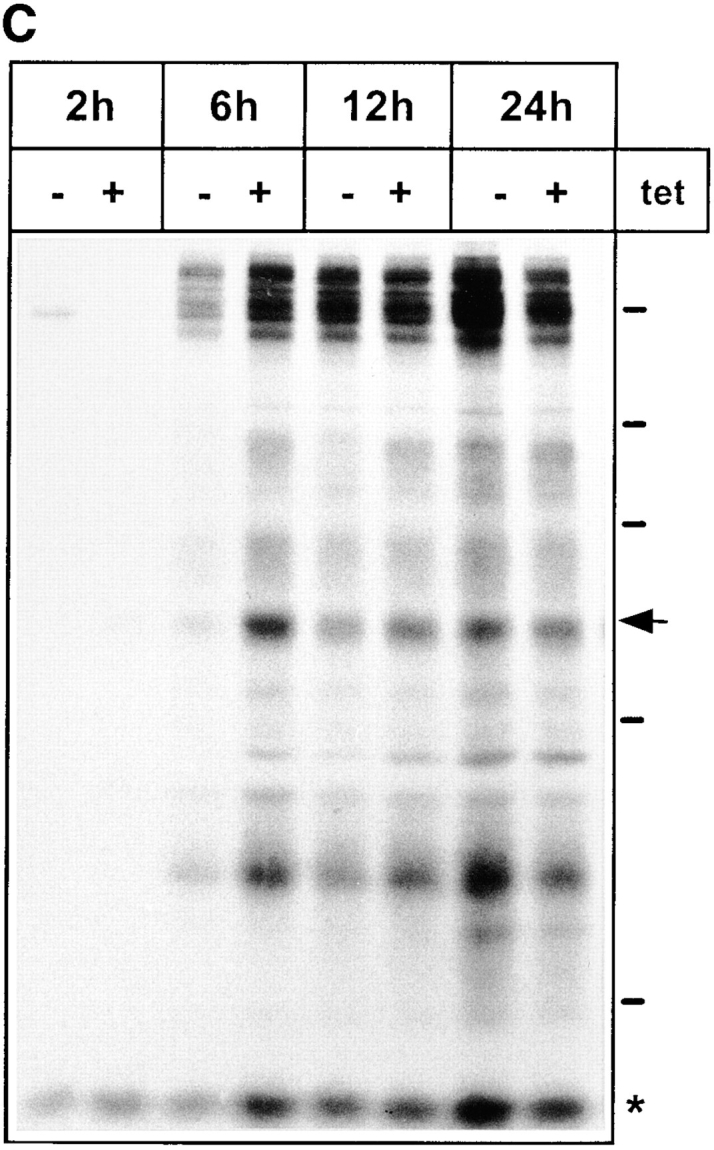
Secretion of glycoproteins in KKFF and KKAA HeLa cells. (A) Cells were incubated for 48 h ± tet, labeled for 30 min with [35S]methionine and chased for 1, 2, and 3 h. Glycoproteins were isolated with Con A beads and separated by 10% SDS-PAGE. 14C-labeled molecular mass markers are indicated at the right margin (200, 97.4, 66, 46, and 30 kD). The arrows indicate the position of the 57-kD protein. *, band used as a reference for quantification in B. The apparent absence of the 66-kD protein in the KKFF panel (3 h, −tet) is due to an artifactual inhomogeneity of the gel. (B) Quantification of the delay in secretion of the 57-kD glycoprotein. The amount of the 57-kD glycoprotein was determined by densitometry of fluorograms and normalized. Shown is the relative secretion as determined by dividing the value obtained for −tet by that for +tet. Values are means ± SEM of at least three independent experiments. white bars, KKFF; black bars, KKAA. (C) Tet- dependent difference in the secretion of the 57-kD protein disappears with increased chase times. KKAA cells were treated ± tet for 48 h, pulsed for 30 min with [35S]methionine, and chased for 2, 6, 12, and 24 h. Glycoproteins were isolated from the culture medium and analyzed by SDS-PAGE as in A.