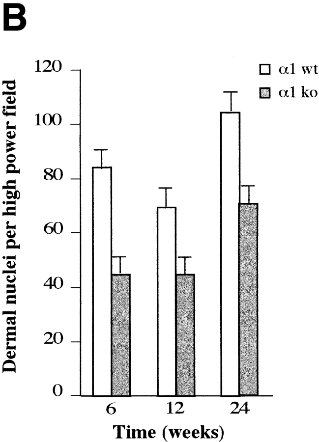
Figure 1.
Fibroblast density in dermis of wild-type and α1-deficient animals. (A) Representative sections of wild-type (WT) and α1-deficient (KO) 6-wk-old skin stained with hematoxylin and eosin. The α1-deficient skin has apparently normal architecture but reduced numbers of nuclei in the dermis. (B) Counts of dermal nuclei in pairs of wild-type and knockout skin obtained from 6-, 12-, and 24-wk-old 129 Sv/ter mice. Each pair of bars represents one pair of apposed sections of back skin sectioned at 5 μm. Note the lower number of dermal nuclei in α1-null dermis compared with that observed in wild-type samples. Bars and errors show mean and standard deviation of counts of dermal nuclei in six high power field views of dermis. Differences between wild-type and α1-null groups were significant with P < 0.05 by Student's t test in each case. Bar, 100 μm.


