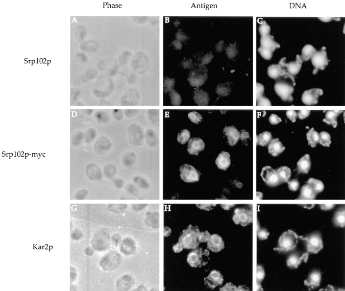
Figure 4.
Immunofluorescent localization of Srp102p to the ER. SOY162 cells containing either pSO454 (Srp102p lacking an epitope tag, A–C and G–I) or pSO457 (Srp102p-myc, D–F) were fixed with formaldehyde and probed with anti-myc (A–F) or anti-Kar2p (G–I) followed by secondary decoration with FITC-conjugated donkey anti-mouse IgG (A–F) or TRITC-conjugated donkey anti–rabbit IgG (G–I). The image in B is exposed for twice the length of time as the image shown in E. Phase contrast images are shown in A, D, and G; fluorescein fluorescence is shown in B and E; rhodamine fluorescence is shown in H, and DAPI staining of the nuclei and mitochondria is shown in C, F, and I.

