
Figure 3.
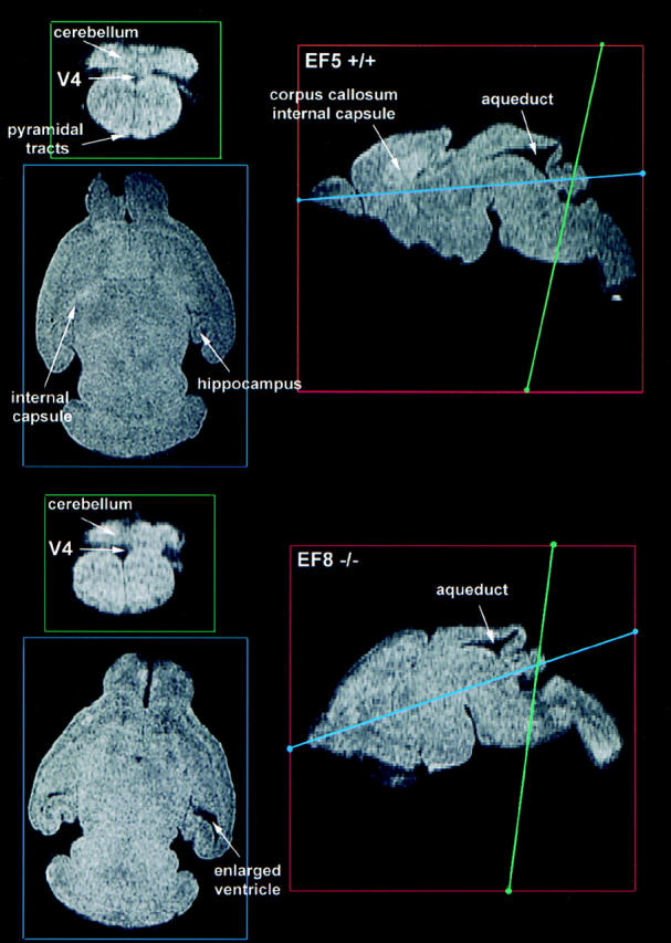
MR microscopy images of brains of normal littermate and ankyrinB (−/−) mice. Parasagittal of brains from a normal littermate (EF5 +/+) and an ankyrinB (−/−) mouse are in red boxes on the right. Sections indicated by blue and green lines are displayed on the left. Although unmyelinated, the internal capsule and pyramidal tracts are clearly visible in the normal brain (top left, blue and green panels) and were not evident in the mutant (bottom left, blue and green panels). The internal capsule and corpus callosum are also visualized in the parasagittal sections of the normal brain (top right, red panel) and could not be resolved in the mutant (bottom right, red panel). The lateral ventricles were abnormally enlarged in mutant brains (bottom left, blue panel). One ankyrinB (−/−) brain exhibited severe malformation of most brain structures and is not shown. These images represent a milder phenotype.