
Figure 4.
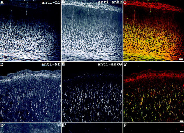
Codistribution of L1 with ankyrinB in premyelinated axon tracts and codistribution of neurofascin with ankyrinG in axon initial segments in neonatal mouse brain. The sections through cerebral cortex of neonatal mouse brain were double labeled with antibodies to L1 (A) and ankyrinB (B, composite image shown in C) or antibodies to neurofascin (D and D′) and ankyrinG (E and E′, composite images shown in F and F′). The results show L1 and ankyrinB codistributed in the axon tracts while neurofascin and ankyrinG colocalized at initial segments of axons. The images shown in D′–F′ were taken from the corpus callosum portion of the same section shown in D–F. Bars, 10 μm.