
Figure 5.
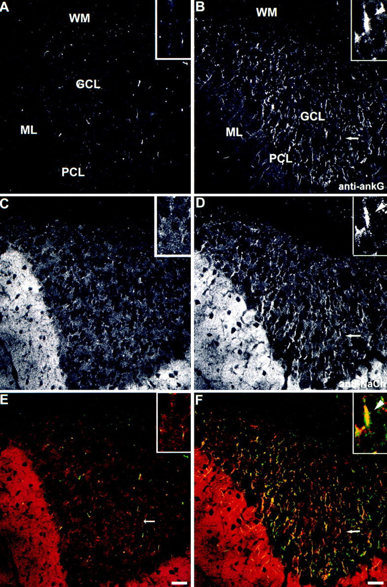
Loss of sodium channel clustering at initial segments of cerebellar granule cell axons of mutant mice. Cerebellar brain sections from mutant mouse (A, C, and E) and the wild-type control littermate (B, D, and F) were double-stained with antibodies to ankyrinG (A and B, green in E and F) and sodium channel (C and D, red in E and F). One typical example of colocalization of ankyrinG and NaCh at the initial segments of granule cells in wild-type mouse cerebellum (B, D, and F, arrows) was magnified and shown in the inset. The initial segment was marked with an arrowhead. Bars, 20 μm.