
Figure 3.
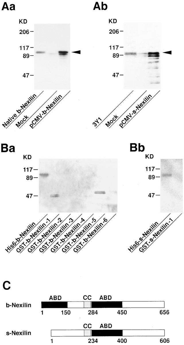
Molecular characterization of b- and s-nexilins. (A) Western blot analysis of recombinant b- and s-nexilins. The pCMV-b- or pCMV-s-nexilin was transfected into COS7 cells, and the cell lysates were subjected to SDS-PAGE (8% polyacrylamide gel), followed by Western blot analysis using the antinexilin antibody. Arrowheads indicate b- and s-nexilins. (Aa) b-Nexilin and (Ab) s-nexilin. (B) 125I-labeled F-actin–binding activity of various truncated forms of b- and s-nexilins. The purified proteins (0.2 μg of protein each), except for GST-b-nexilin-1, were subjected to SDS-PAGE (10% polyacrylamide gel), followed by 125I-labeled F-actin blot overlay. GST-b-nexilin-1 was recovered in the inclusion bodies during the preparation of the supernatant of the cell lysates, and thus an aliquot of the cell lysates was used for the assay. GST-b-Nexilin-1, aa 1–150; GST-b-Nexilin-2, aa 77– 240; GST-b-Nexilin-3, aa 151–283; GST-b-Nexilin-4, aa 211–330; GST-b-Nexilin-5, aa 284–450; GST-b-Nexilin-6, aa 493–656; and GST-s-Nexilin-1, aa 1–100. (Ba) b-Nexilin and (Bb) s-nexilin. (C) Schematic drawing of the structures of b- and s-nexilins. ABD, F-actin–binding domain; CC, coiled-coil region.