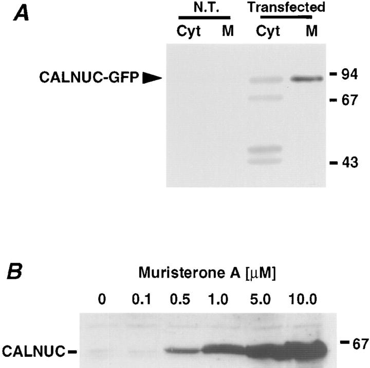
Figure 2.
Biochemical analysis of CALNUC expression in transfected cells. (A) HeLa cells transiently transfected with CALNUC-GFP. By immunoblotting (50 μg protein), ∼85% of the CALNUC-GFP (90 kD) is associated with the membrane pellet (M), and the remaining 15% is found in the cytosolic fraction (Cyt). No signal was detected in nontransfected HeLa cells (N.T.) when the same amount of protein was examined. HeLa cells transfected with CALNUC-GFP cDNA (48 h) were homogenized and membrane (100,000 g pellet) and cytosolic (100,000 g supernatant) fractions were prepared from the postnuclear supernatant. The pellet was resuspended in the same volume as the supernatant, and equal volumes of the membrane and cytosolic fractions were analyzed by SDS-PAGE and immunoblotted with affinity-purified anti-CALNUC IgG. (B) Expression of CALNUC in inducible, stably transfected EcR-CHO-CALNUC cells. By immunoblotting increasing amounts of CALNUC were detected in cell lysates (80 μg) after induction with increasing amounts (0.5–10 μM) of muristerone A. Cells stably transfected with CALNUC were lysed in 1% Triton X-100. Lysates (80 μg) were separated by SDS-PAGE and immunoblotted with affinity-purified anti-CALNUC IgG.

