
Figure 1.
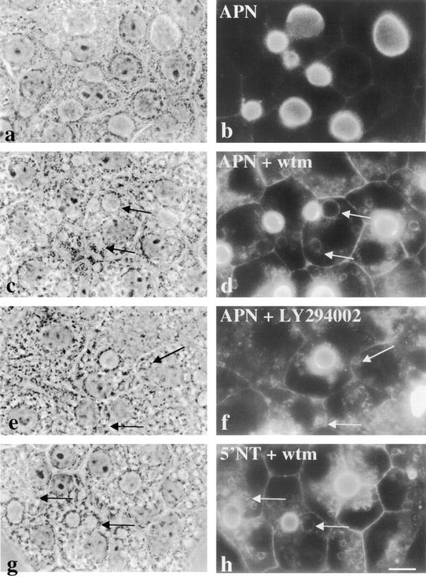
Apical plasma membrane proteins accumulate in vacuoles in the presence of wortmannin (wtm) or LY294002. WIF-B cells were incubated for 180 min in the absence (a and b) or presence (c, d, g, and h) of 100 nM wortmannin or 200 μM LY294002 (e and f). Cells were fixed, permeabilized and stained for APN (b, d, and f) or 5′NT (h). Arrows point to vacuoles containing intracellular apical proteins. Fluorescent images were intentionally overexposed so that vacuolar staining was more visible. The control staining in b was overexposed to the same extent as treated cells. 5′NT staining in control cells is indistinguishable from that of APN shown in a. Bar, 10 μm.