
Figure 5.
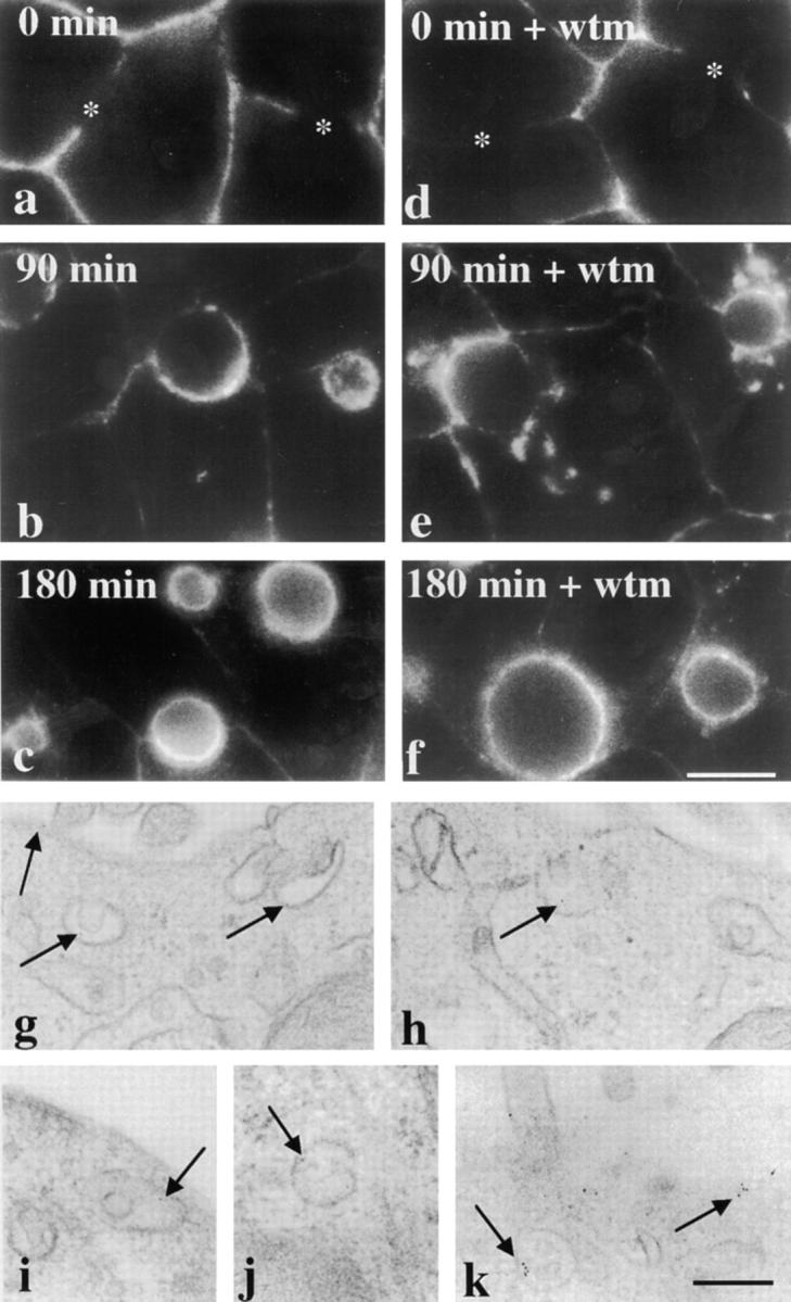
Transcytosing apical plasma membrane proteins bypass vacuoles. (a–f) Cells were pretreated for 15 min in the absence or presence of 100 nM wortmannin (wtm) at 37°C. The cells were chilled to 4°C and labeled with anti–5′NT antibodies for 15 min. After extensive washing, the cells were either fixed and permeabilized directly (0 min of chase; a and d) or placed at 37°C and the antibodies chased for 90 (b and e) or 180 (c and f) min in the continued absence or presence of 100 nM wortmannin. After chase, the cells were fixed, permeabilized, and the trafficked antibodies detected with cy3-conjugated secondary antibodies. The control experiment is shown in a–c and wortmannin-treated cells are shown in d–f. Asterisks in a and d point to unlabeled bile canalicular domains at 0 min of chase in both control and treated cells. Bar, 10 μm. (g–k) Cells were pretreated and labeled as described above, except anti–5′NT antibodies were directly conjugated to 5-nm gold particles. The antibodies were chased for 90 min in the continued absence (g and h) or presence (i–k) of 100 nM wortmannin and processed for electron microscopic visualization (see Methods). Arrows are pointing to gold particles located at the apical cell surface or in the SAC in both control and treated cells. Bar, 250 nm.