
Figure 3.
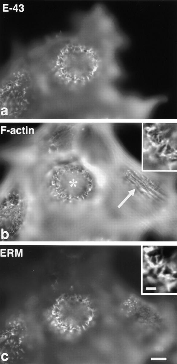
Changes in cortical actin filament organization in L fibroblasts transiently overexpressing E-43. Cells were triply stained with anti–E-cadherin mAb (E-43, a), FITC-phalloidin (F-actin, b), and anti-ERM mAb (ERM, c). Nontransfected cells (compare a with b) were characterized by dorsal stress fibers (arrow in b) and short microvilli (c), whereas in E-43–overexpressing cells long microvillar core actin bundles were induced with concomitant disruption of dorsal stress fibers (asterisk in b). The precise colocalization of F-actin and ERM proteins in elongated microvilli in E-43–expressing cells is shown at higher magnification in the insets. Cells were transfected by microinjection. Bar, a–c, 10 μm; insets, 2 μm.