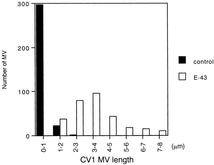
Figure 5.
Length distribution of microvilli in E-43–expressing (open box) and nontransfected (closed box) CV-1 cells. The length of each microvillus was measured from anti-ERM mAb-stained images (see Fig. 4 b). In nontransfected cells, microvilli were relatively short (0.53 ± 0.33 μm; n = 320), whereas in E-43– overexpressing cells they were significantly elongated (3.5 ± 1.4 μm; n = 300). Cells were transfected by lipofection.

