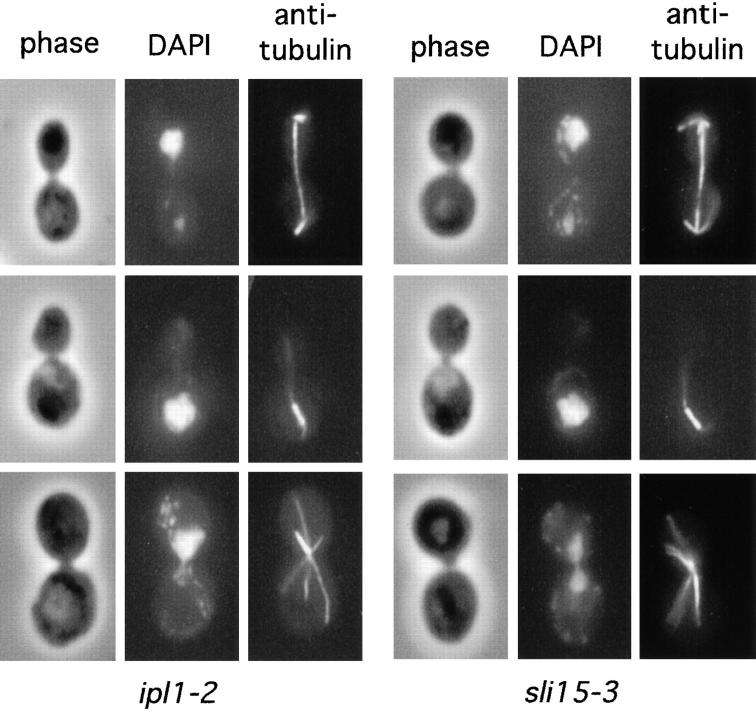
Figure 1.
Cytological phenotype of ipl1-2 and sli15-3 mutants. ipl1-2 (CCY108-15C-1) and sli15-3 (CCY1060-1D) cells were incubated at 37°C for 4 h and processed for immunofluorescence microscopy. Phase-contrast, DAPI-stained, and anti-tubulin– stained images are shown. Top panels: uneven chromosome segregation; middle panels: nuclear migration defect; bottom panels: monopolar spindle.

