
Figure 6.
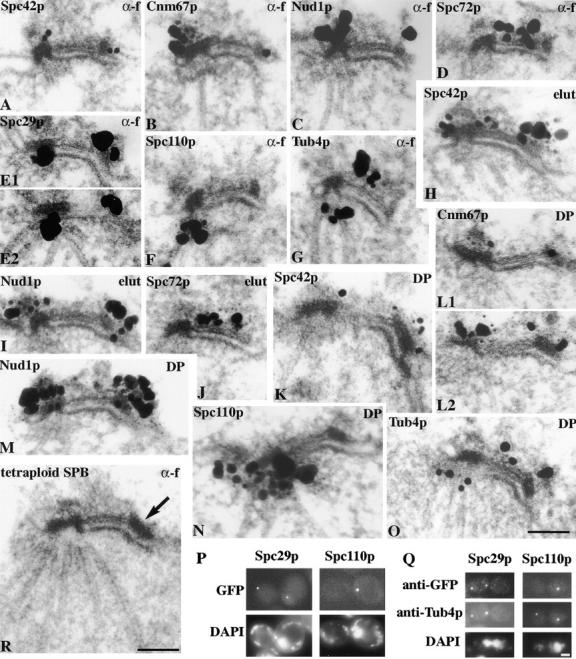
ImmunoEM localization of GFP-labeled Spc42p, Cnm67p, Nud1p, Spc72p, Spc29p, and Spc110p in cells arrested in G1 with α-factor (G1 α-f), G1 elutriated cells grown for 25 min (elut), and cells released from α-factor for 30 min to observe the duplication plaque (DP). Tub4p was detected with affinity-purified polyclonal antibodies. Adjacent serial sections have the same panel letter and are numbered. (P) Fluorescence of GFP-Spc29p and GFP-Spc110p in unfixed mps2 cells after 3 h at 37°C then treated with DAPI for 0.5 h at 37°C; (Q) immunofluorescence of the same cells with anti-GFP and anti-Tub4p; (R) EM of a thin section of a tetraploid MATa cell arrested with α-factor. The arrow indicates the satellite. Bars, 0.1 μm for the immunoEM and EM and 2 μm for the light microscopy.