
Figure 1.
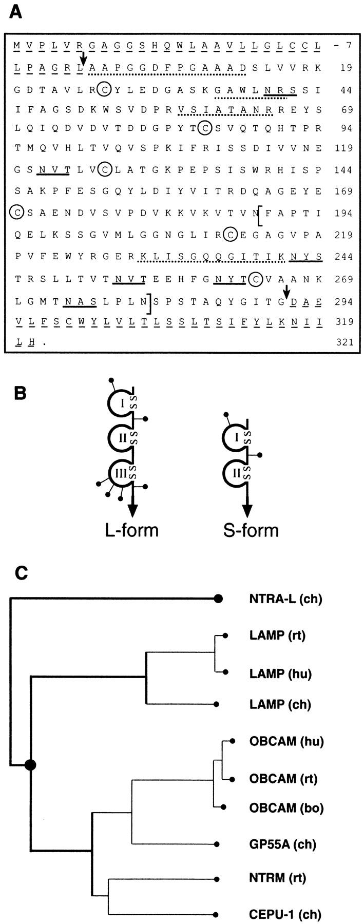
Primary structure, domain models of neurotractin and sequence relationship to other IgLON members. (A) Primary structure of neurotractin. The predicted NH2-terminal signal peptide and the COOH-terminal hydrophobic segment are underlined by dashed lines and arrows indicate the mature NH2 terminus and COOH terminus. Putative N-linked glycosylation sites are underlined and characteristic cysteine residues of Ig-like domains are labeled by circles. To obtain independent evidence that the protein which had been isolated by immunoaffinity chromatography (see Fig. 2 B, lane 1) is identical with that predicted by the cDNA clones, a sample of it as well as peptides derived from a tryptic digest were subjected to Edman degradation. Evaluation of the partial internal as well as the NH2-terminal sequence (indicated by dotted lines) confirms that they match this cDNA sequence. The alternatively spliced and L-form–specific third Ig-like domain is indicated by brackets. The sequences of S-form and L-form are available from GenBank/EMBL/DDBJ under accession numbers AJ132998 and AJ132999, respectively. (B) Domain models of neurotractin-L (large) and -S (small). Ig-like domains are drawn as loops that are closed by disulfide bridges, putative N-linked glycosylation sites are shown as lines ending with dots and the GPI-anchor is represented by an arrow. (C) Neurotractin is a novel protein belonging to the IgLON subgroup. Sequence relationship of neurotractin-L to other members of the IgLON subgroup was examined with the PILEUP program from the GCG package (University of Wisconsin, Madison, WI) and sequences have been taken from Schofield et al., 1989; Lippman et al., 1992; Shark and Lee, 1995; Struyk et al., 1995; Pimenta et al., 1995, 1996a; Spaltmann and Brümmendorf, 1996; Brümmendorf et al., 1997; Hancox et al., 1997. GP55-A is a partial sequence lacking most likely a short stretch at the NH2 terminus (Wilson et al., 1996). ch, chicken; hu, human; bo, bovine; rt, rat.