
Figure 3.
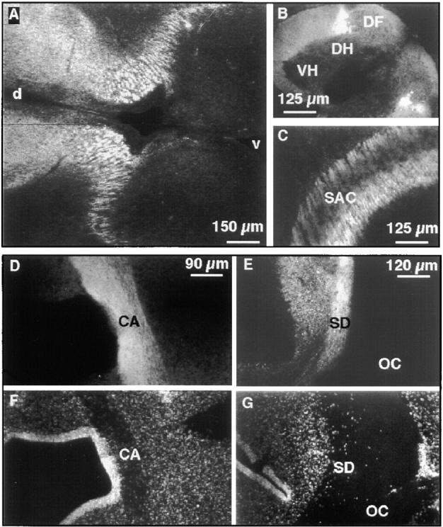
Neurotractin expression on subsets of embryonic axon tracts. Immunohistochemical analysis of E7 diencephalon with mAb NTRA-1 shows crosscut longitudinal neurotractin positive axon tracts (A). In the E9 spinal cord dorsolateral longitudinal axons show strong neurotractin expression (B). In tectum of embryonic day 8 neurotractin is found on axons of the stratum album centrale (C). Staining of a horizontal section of E9 ventral forebrain with mAb NTRA-1 shows strong expression of neurotractin on axons of the anterior commissure (D). This axon tract is also discernible as a nuclei-poor zone as revealed by DNA staining (F). A horizontal section at the level of the E9 optic chiasm reveals neurotractin expression on axons of the supraoptic decussation (E). Bisbenzimide staining of cellular nuclei in the same section outlines the axons of the supraoptic decussation as well as those of retinal ganglion cells in the optic chiasm as an extended nuclei-poor region (G). Note that neurotractin is restricted to the supraoptic decussation and is lacking on the retinal ganglion cell axons. At E9, the supraoptic decussation is not yet subdivided into the dorsal, ventral and subventral regions which can be distinguished later in development (Ehrlich et al., 1988). VH, ventral horn; DH, dorsal horn; DF, dorsal funiculus; SAC, stratum album centrale; d, dorsal; v, ventral; CA, commissura anterior; SD, supraoptic decussation; OC, optic chiasm.