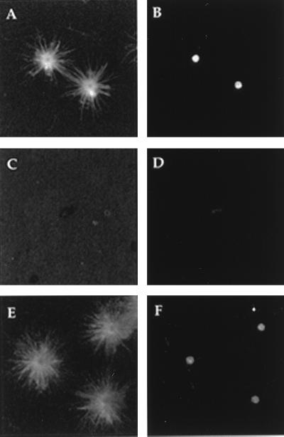
Figure 1.
Fluorescence micrographs of asters formed by centrosomes and centrosome remnants after indirect immunofluorescent labeling for both tubulin (A, C, and E) and γ-tubulin (B, D, and F). The MNP (A) and γ-tubulin (B) present in centrosomes are removed by treatment with 1.0 M KI (C and D). The MNP recovers (E) and γ-tubulin returns (F) when KI-insoluble centrosome remnants are incubated in oocyte extracts.

