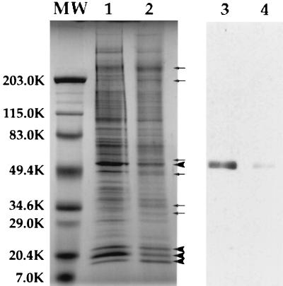
Figure 2.
SDS/PAGE and immunoblot analyses of centrosomes and KICRs. Some of the proteins present in isolated centrosome fractions (lane 1) are removed by KI treatment (lane 2). Three proteins of ≈20 kDa, which copurify with centrosomes (lane 1), along with a protein of ≈50 kDa are the most abundant proteins in KICR fractions (large arrowheads, lane 2). Additionally, at least six proteins are enriched in the KICR fraction compared with control centrosomes (small arrows, lane 2). Immunoblots of centrosomes (lane 3) and KICRs (lane 4) with affinity-purified polyclonal anti-γ-tubulin antibody reveals that γ-tubulin that is present in isolated centrosome fractions (lane 3), is removed by KI treatment and KICR fractions (lane 4) contain negligible γ-tubulin.

