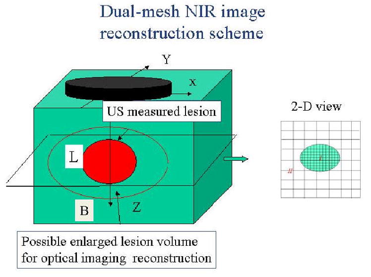
Fig.4.

Illustration of the dual-mesh optical imaging reconstruction with the assistance of ultrasound lesion mapping. The entire imaging volume is segmented into lesion (L) and background regions (B). The finer imaging grid is used for the lesion region and the coarse grid is used for the background. To account for possible larger angiogenesis extension of the ultrasound-identified lesions, we have used a much larger region of interest (ROI) than the US-identified one for finer grid lesion mapping. In general, an elliptical ROI is used in optical imaging reconstruction.
