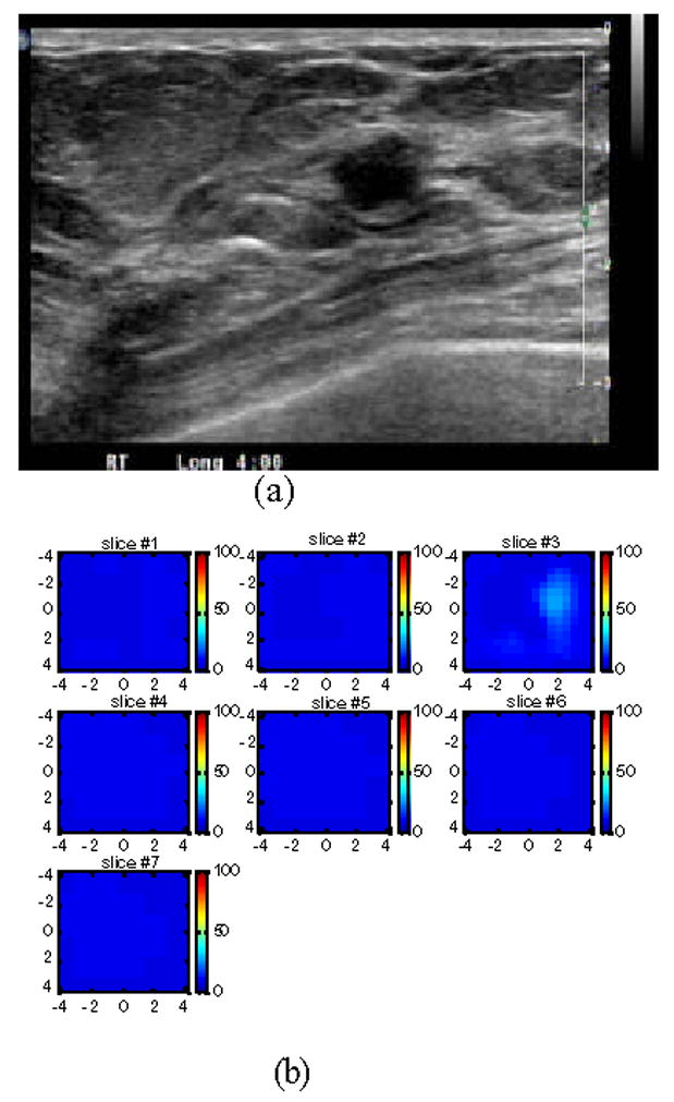
Fig.6.

(a) US image of a suspicious solid lesion measured 8.4 mm × 6 mm located at 4–5 o’clock of the right breast of a 38-year old woman. The biopsy result revealed that the lesion was a fibroademona with no evidence of malignancy. (b) The total hemoglobin concentration map computed from absorption maps of 780nm and 830nm (not shown). No isolated mass with high blood content is seen. The vertical scale is the total hemoglobin concentration in μ mol ranging from 0 to 150 μ mol/liter.
