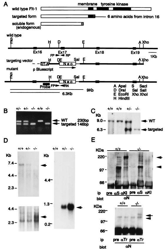
Figure 1.
Targeted inactivation of the tyrosine kinase domain of Flt-1 in ES cells and mice. (A) Predicted structures of wild-type, targeted, and endogenously expressed soluble Flt-1 (Upper), as well as the restriction maps of flt-1 wild-type allele, replacement-type targeting vector, and the targeted allele after homologous recombination (Lower). The targeting vector contains a PGK-neo cassette (pPGKneopA) and the diphtheria toxin A gene in the upstream region of the construct. (B and C) Genotype of a litter from flt-1TK+/− and flt-1TK+/− interbreeding. Tail DNA was isolated and analyzed by PCR (B) and Southern blotting (C) by using the probe shown in A after digestion with EcoRI. Primers used also are shown in A as FP (forward primer), RP (reverse primer), and RN (reverse neo primer). (D) Northern blot analysis of poly(A) RNAs (3 μg/lane) extracted from wild-type, flt-1TK+/−, and flt-1TK−/− mice of whole embryos. Probes used were the 3′ half of mouse flt-1 cDNA (nucleotide residue 2643–4008) containing the tyrosine kinase domain (Upper Left) and the 5′ half of flt-1 cDNA (69–1919) (Lower Left) and neo sequence (Right). The arrow in the upper left indicates the full-length mouse flt-1 mRNA of 7.0 kb long. (E) Immunoblot analysis of proteins obtained from mouse lung with various antibodies (Ab). pre, preimmune rabbit serum; aS, Ab against the soluble Flt-1 specific for the carboxyl terminal 31 amino acids (arrowhead, Upper); aKI, Ab against the kinase insert that is encoded from exon 21 (arrow); aN, Ab against the amino terminal region of the Flt-1 extracellular domain; aTr, Ab specific to the carboxyl terminal sequence of truncated Flt-1 (arrow, Lower). Arrowheads show nonspecific bands (Lower).

