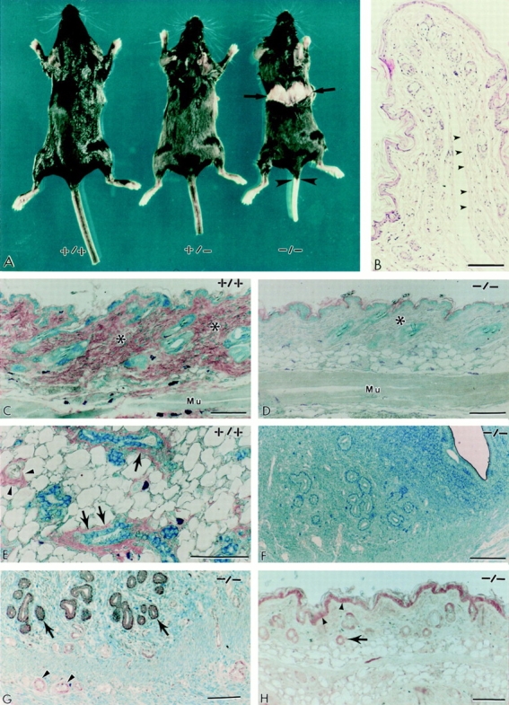
Figure 3.

The phenotype of decorin-deficient mice reveals thinning and fragility of the skin. (A) Gross photograph of wild-type (+/+), heterozygous (+/−), and homozygous (−/−) littermates. Notice the sharp rupture of the back skin in the −/− animal (arrows) and the detachment of the tail skin (arrowheads) that occurred during cervical dislocation. The skin fragility was never observed in either +/+ or +/− animals (n > 300). B is a cross-section of the detached tail skin. Notice the sharp and bloodless line of rupture (arrowheads) along the deeper dermis. Immunohistochemical analysis of skin and skeletal muscle (C and D), mammary gland (E), and uterus (F) from wild-type (+/+) and decorin-deficient (−/−) animals using the LF-113 anti-decorin antibody. Notice the intense immunoreactivity in the dermis of a wild-type animal (C, asterisks) and the fine immunoreactivity on either side of the skeletal muscle (Mu). In contrast, the −/− dermis (D, asterisk), the subcutaneous and perimysial connective tissues, are totally unreactive as are the uterine wall and mucosa (F). As expected, the anti-decorin antibody labeled specifically the adventitia of small blood vessels (E, arrowheads) and the myoepithelial cells and fine connective tissue surrounding mammary ducts (E, arrows). Immunodetection of biglycan using an anti-peptide (LF-106) antibody in uterus (G) and skin (H) from Dcn−/− mice. Notice the “normal” expression of biglycan in the endometrium (G, arrows) and in the intramural small blood vessels (G, arrowheads). As expected, in skin, both the epidermis (H, arrowheads) and the follicular epithelium (H, arrow) were labeled by the anti-biglycan antibodies. Sections were reacted with LF-113 or LF-106 antisera at 1: 1,000 dilution, and then visualized with peroxidase-conjugated IgG (1:200) followed by counterstaining with 0.2% methylene blue. Bar, 100 μm.
