
Figure 5.
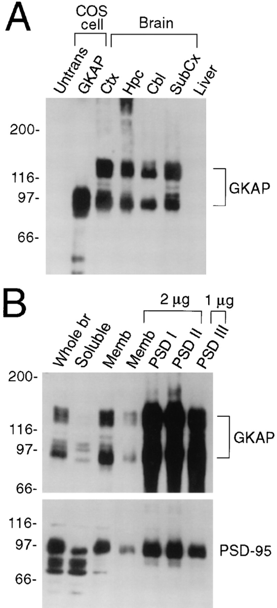
Expression pattern of GKAP protein in rat brain. (A) Specificity of GKAP antibodies and differential regional expression of GKAP in rat brain. Whole cell extracts of untransfected COS-7 cells (Untrans.), or of COS cells transfected with GKAP cDNA, were analyzed by immunoblotting with GKAP2.1 antibodies, along with membrane fractions (10 μg protein) from different regions of brain or liver, as indicated. Ctx (cortex), Hpc (hippocampus), Cbl (cerebellum), Subcx (subcortical regions). Positions of molecular size markers are shown in kD. (B) Immunoblot analysis of subcellular fractionation of GKAP. Lanes were loaded with rat brain fractions, as follows: Whole br (total brain homogenate, 20 μg protein), Soluble (S100 supernatant fraction of brain homogenate, 30 μg). Memb (crude synaptosomal membrane fraction, 10 μg or 2 μg, as indicated); PSDI, PSDII, and PSDIII (purified PSD fractions after extraction with Triton X-100 once [I], twice [II], or with Triton X-100 followed by sarkosyl [III]). Filters were probed with GKAP and PSD-95 antibodies, as indicated. (Equal percentages [rather than equal mass] of membrane and soluble fractions were loaded—the soluble fraction contained three times higher concentration of total protein than the membrane fraction. To show relative purification in the PSD fractions, only 2 μg of PSDI, PSDII, and 1 μg of PSDIII were immunoblotted and compared with 2 μg of synaptosomal membrane fraction.)