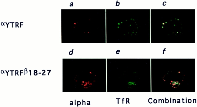
Figure 8.
Localization after endocytosis of αY or αYβ18–27 relative to transferrin receptor. K562 cells stably transfected with αY (top) or αYβ18–27 (bottom) were incubated at 37°C for 2 h with anti-α mAb 7G7B6. The cells were then washed at 4°C to stop endocytosis, fixed, permeabilized and incubated with anti-transferrin receptor mAb OKT9. Anti-α and antitransferrin receptor mAb were then revealed with Texas red– labeled anti–mouse IgG2a and FITC labeled anti–mouse IgG1, respectively. One representative medial optical section is represented. (a and d) α staining; (b and e) transferrin receptor staining; and (c and f) combinations of the two stainings.

